
News ·
BliXray applies composite and powder CT to Europe's ceramic matrix composite scale-up
BliXray offers Laboratory CT analysis of fibre-reinforced composites and powders to producers scaling up ceramic matrix composite parts. The CT data show fibre direction, fibre distribution and particle structure in 3D, without cutting the sample.
Computed tomography (CT) reconstructs a sample as a three-dimensional volume. The internal architecture of a composite is then measured rather than inferred from a polished section. For a materials engineer qualifying a new composite, this means one non-destructive data set per sample. The sample itself stays available for mechanical testing or further processing.
Fibre architecture measured in three dimensions
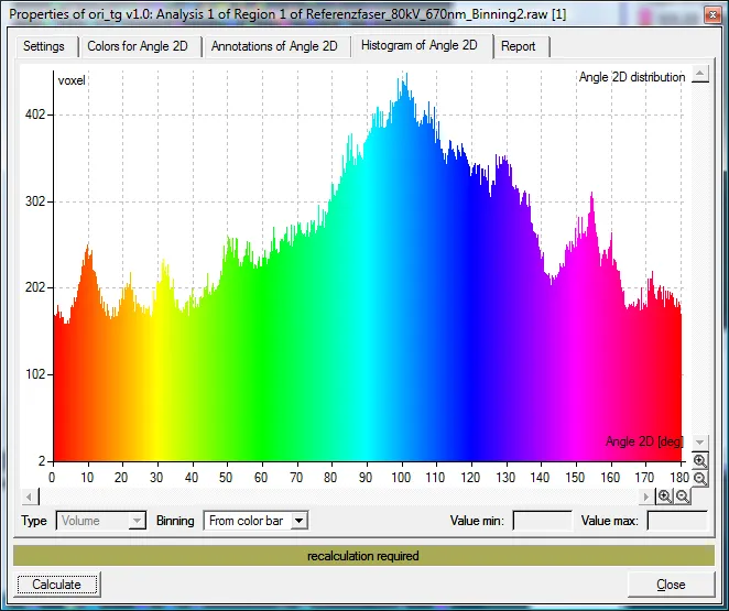
The behaviour of a fibre-reinforced part depends on where its fibres point and how evenly they are spread. BliXray’s Laboratory CT analysis has been shown on short-fibre and carbon-fibre reinforced polymers (SFRP and CFRP). From the reconstructed volume, the analysis software returns:
- fibre direction through the sample;
- fibre concentration and its local variation;
- fibre distribution across the full volume.
Fibre misalignment and local changes in fibre content appear as regions in the volume, not as a single average value. The engineer can locate them, compare them between batches and relate them to process settings. Porosity that concentrates stress is recorded in the same data set.
“A cross-section tells you about one plane of a composite. A CT volume tells you about all of them,” says Adrian de Riz, Managing Director, BliXray Technologies GmbH. “Our customers get fibre orientation and distribution as data they can compare from batch to batch.”
Powder and particle data from the same instrument
Many ceramic and composite process routes begin with a powder, and the quality of the final part starts there. The same Laboratory CT analysis calculates particle count, surface area, volume, sphericity and compactness for powder and particle samples. Because these values come from a 3D volume, they describe particle shape in every direction rather than a projected outline. Particle-size and particle-shape distributions can therefore be followed from one powder lot to the next. For a powder supplier or a part maker, a shape question becomes a measured value per lot. The method has also been applied qualitatively to a carbide specimen, a dense and hard material.
Ceramic matrices treated as a test case
Ceramic matrix composites raise the same questions as polymer composites: fibre direction, fibre distribution and porosity. The matrix is denser, however, and this changes the contrast between fibre and matrix. BliXray therefore treats every new ceramic matrix material as a test case. A test scan of the customer’s own sample shows which features the analysis resolves before routine use is agreed. Where a feature sits close to the limit of what the scan shows, the result says so rather than reporting it as found. Scan settings are then chosen for the ceramic material itself, not carried over from a polymer composite.
For aerospace and defence supply chains, the result is a documented, non-destructive record of each qualification sample. Development laboratories and research groups use the same data to link process changes to the internal structure they produce.
“We scan the customer’s own material first and show what the data can and cannot tell them,” says de Riz. “That gives a materials team a sound basis before it builds CT into its qualification routine.”
Test scans available
BliXray offers test scans of composite, ceramic and powder samples and discusses the fibre and particle data with the customer’s materials team. Contact Adrian de Riz, Managing Director, BliXray Technologies GmbH, a.deriz@blix-ray.de, +49 1578 0477753 – www.blix-ray.de.
About BliXray – BliXray Technologies GmbH, Hannover, designs and builds industrial CT systems and X-ray safety cabinets. Manufacturers in defence, aerospace and research use BliXray CT to inspect safety-critical parts without destroying them. BliXray cabinets and containers house X-ray sources up to 300 kV at a protection level of 0.5 µSv/h.